

Precision root canal therapy with microscopic techniques
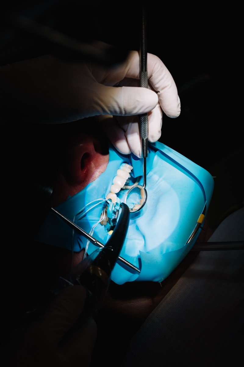
You have likely heard the stories—or perhaps lived through one yourself. You visit a dentist for a toothache, undergo a procedure, and think the problem is solved, only for the pain to return months or years later. It is a frustrating cycle that leads many patients to believe that tooth extraction is the only permanent solution. However, in the vast majority of these cases, the failure isn’t due to the biology of your tooth, but rather the limitations of human vision.
When you are facing a complex diagnosis or the prospect of re-treating a previously worked-on tooth, the technology used during your procedure is just as critical as the clinician’s hands. For patients seeking expert dental care in South Kensington, the integration of high-powered microscopic endodontics is changing the narrative. It transforms what was once a “blind” procedure into a precise, predictable science, offering a second chance to teeth that might otherwise be lost.
The Invisible Challenge: Why Traditional Methods Struggle
To understand why microscopic dentistry is a game-changer, we first need to rethink the anatomy of a tooth. We often imagine a root canal as a simple, straight pipe—like a straw. In reality, the root system of a tooth is more like a complex cave system or a river delta. It twists, turns, branches off into tiny tributaries, and narrows into spaces thinner than a human hair.
For decades, dentists performed root canal therapy primarily by “feel” and with the naked eye. While effective for simple cases, this approach has inherent limitations:
- Shadows: The interior of a tooth is dark. Traditional dental lights cannot illuminate the deepest curves of a root system.
- Hidden Anatomy: If a dentist cannot see a canal, they cannot clean it. Bacteria left behind in these hidden spaces are the primary cause of treatment failure.
- Structural Integrity: Without magnification, a dentist often has to remove more tooth structure to find the canals, potentially weakening the tooth.
This is where the difference between “good enough” and “precision” becomes clear.
The Microscopic Difference: Seeing the Unseen
At Behrens Dental Practice, we believe that you cannot treat what you cannot see. This is why we utilize high-powered dental operating microscopes for endodontic procedures. This isn’t simply a magnifying glass; it is a sophisticated optical system that provides up to 25x magnification and co-axial illumination (meaning the light goes exactly where the eyes look, eliminating shadows).
For a patient requiring root canal treatment in London, this technology shifts the experience from one of uncertainty to one of precision. The microscope allows the dentist to look directly down into the root channels, identifying issues that would be completely invisible to the naked eye.
The “Aha” Moment: How Precision Solves the 3 Biggest Problems
If you have been referred for a complex root canal, it likely falls into one of three categories. Here is how microscopic endodontics addresses each one specifically.
1. The Mystery of the “MB2” Canal
The most common reason for root canal failure in upper molars is a missed canal, specifically known as the “Mesiobuccal 2” (MB2) canal. Research suggests that while this fourth canal is present in over 90% of first molars, it is frequently missed during traditional treatment because it is exceptionally tiny and often hidden under a ridge of dentin.
With a microscope, we can spot the microscopic “trough” that indicates where this hidden canal lies. By locating and cleaning this fourth canal, we dramatically reduce the risk of persistent infection.
2. Navigating Calcified Canals
As we age, or in response to past trauma, our teeth naturally deposit more hard tissue (dentin), causing the nerve space to shrink. These are called “calcified canals.” To the naked eye, it looks like the canal has disappeared entirely.
Without magnification, a dentist might attempt to drill down to find the canal, risking perforation of the root. With microscopic guidance, we can visually track the minute change in color that marks the path of the original canal, allowing us to negotiate these narrow passages safely and preserve the tooth.
3. Retreatments and Broken Instruments
Sometimes, a previous root canal tool (a file) may have separated inside the tooth during a past procedure, blocking the canal. Alternatively, old filling material needs to be removed carefully to treat a new infection. The microscope allows for the precise removal of these obstructions without damaging the surrounding wall of the root—a feat that is nearly impossible to perform safely with the naked eye.
The Evidence: Data-Driven Confidence
It is natural to wonder if this technology actually translates to better results. The data is compelling. Studies focusing on complex cases—specifically molars—have shown that microscope-assisted root canal therapy significantly increases the probability of a successful outcome.
In fact, research indicates that the odds of success can be 2.9 to 3.2 times higher when using a microscope compared to traditional methods. This isn’t just about clinical success; it’s about your peace of mind. Precision means:
- Less Trauma: Because we can see exactly where to go, we don’t need to remove unnecessary tooth structure.
- Fewer Appointments: We can often locate and treat all canals in a single visit, rather than bringing you back multiple times to “wait and see.”
- Reduced Discomfort: Precise treatment leads to less post-operative inflammation.
Beyond the Procedure: Protecting Your Investment
Saving a tooth through precision endodontics is a victory for your oral health, but it is part of a larger ecosystem. Once a tooth has been restored, maintaining the health of the surrounding gums and bone is vital to ensure that the tooth remains stable for life.
Many patients who struggle with root issues also have underlying gum concerns. Integrating your recovery with proper periodontal maintenance ensures that the foundation holding your newly saved tooth remains strong. It is this holistic approach—combining restorative precision with preventative care—that defines the Behrens philosophy.
FAQ: Common Questions About Microscopic Endodontics
Q: Is a microscopic root canal more painful than a regular one?
A: No, quite the opposite. Because the microscope allows for extreme precision, the procedure is less invasive. We target only the infected tissue, sparing healthy tooth structure, which typically results in less post-operative tenderness.
Q: My dentist said my canals are “too curved” to fix. Can a microscope help?
A: Often, yes. Curved canals present a challenge because “blind” instruments can’t navigate the bend without damaging the root. The visibility provided by the microscope, combined with modern flexible nickel-titanium files, allows us to navigate complex curvatures that were previously considered untreatable.
Q: Does this take longer than a standard appointment?
A: While the setup requires expertise, the procedure is often more efficient. Because we aren’t “searching in the dark” for canals, we can locate and clean them faster. This efficiency often prevents the need for multiple visits.
The Next Step in Your Journey
If you have been told you have a “difficult” tooth, or if you are experiencing pain in a tooth that has already been treated, do not assume extraction is the only option. The difference between losing a tooth and keeping it for a lifetime often comes down to the precision of the diagnosis.
At Behrens Dental Practice in South Kensington, we combine heritage with cutting-edge technology to ensure every patient understands their options. We invite you to experience the difference that precision, transparency, and advanced care can make for your smile.